
Fifty Years of GHK-Cu Research
Loren Pickart didn't set out to discover an anti-ageing compound. He was studying why young plasma extended the lifespan of ageing liver cells in culture — a classic problem in cell biology — and what he found in 1973 was a small tripeptide, tightly bound to a copper ion, that appeared to drive much of the regenerative effect. The compound was Glycyl-L-Histidyl-L-Lysine copper(II), or GHK-Cu. Pickart published his findings in Nature (1973), and then essentially watched the scientific mainstream walk past them for two decades before the compound started attracting serious attention.
Fifty years later, a new in vitro study from a European dermatology research group has added fresh data to GHK-Cu's growing research profile — specifically examining its effects on collagen synthesis pathways in human dermal fibroblasts. The findings confirm some of what was already suspected and add mechanistic detail that earlier studies couldn't provide.
The Proposed Mechanisms of GHK-Cu
GHK-Cu is interesting partly because it's active at genuinely low concentrations — peak biological activity in cell culture systems occurs around 1 nM, with effects declining above 1 μM. That's a picomolar-to-nanomolar activity range, consistent with a compound acting through receptor-like interactions rather than bulk biochemistry.
TGF-β/Smad Signalling
GHK-Cu upregulates TGF-β1 (transforming growth factor beta-1) autocrine signalling, leading to Smad2/3 phosphorylation and transcriptional activation of COL1A1 and COL3A1 genes. This is the primary collagen synthesis mechanism confirmed in the new fibroblast study.
MMP-1 Suppression
Matrix metalloproteinase-1 (collagenase) expression is suppressed by GHK-Cu at the same concentration range that stimulates collagen synthesis. MMP-1 is the primary enzyme that degrades existing collagen fibres — its suppression both protects existing matrix and amplifies net collagen accumulation.
VEGF and Hair Follicle Effects
GHK-Cu stimulates VEGF (vascular endothelial growth factor) and KGF (keratinocyte growth factor) production in dermal papilla cells, which are associated with hair follicle growth regulation. This is the basis for GHK-Cu's interest in hair biology research.
Anti-Inflammatory Signalling
GHK-Cu suppresses NF-κB pathway activation and reduces pro-inflammatory cytokine expression (IL-6, TNF-α) in dermal cell cultures. This anti-inflammatory activity may be synergistic with the collagen-stimulating effects in wound healing research contexts.
What the New Fibroblast Study Found
The study used primary human dermal fibroblasts from five donors (passage 4–6), treated with GHK-Cu at concentrations ranging from 0.1 nM to 10 μM. The primary readouts were COL1A1 and COL3A1 mRNA expression (qRT-PCR), collagen protein secretion (hydroxyproline assay), MMP-1 expression, and TGF-β1 pathway activity markers (pSmad2/3, TGF-β1 secretion ELISA).
The key findings: collagen synthesis peaked at approximately 1 nM GHK-Cu, with 2.3-fold COL1A1 and 1.8-fold COL3A1 mRNA upregulation over untreated controls. MMP-1 expression was suppressed by approximately 25–30% across the same concentration range. TGF-β1 secretion and pSmad2/3 levels were both elevated, confirming TGF-β autocrine signalling as a central mechanism. Critically, the study confirmed the biphasic dose-response: at concentrations above 1 μM, the collagen-stimulating effect diminished, and two of the five donor cell lines showed collagen synthesis below control levels at 10 μM.
The donor variability was also notable. Effect sizes ranged from approximately 1.8-fold to 2.8-fold COL1A1 upregulation at peak concentration across the five donors. This is relevant for researchers designing in vitro experiments — GHK-Cu's effects are real and consistent in direction, but not uniform in magnitude across different primary cell cultures.
Key Finding: Biphasic Dose-Response Confirmed
The hormetic dose-response of GHK-Cu — activity peaking around 1 nM and declining at higher concentrations — is now confirmed by this five-donor primary cell study and is important for in vitro research design. Using GHK-Cu at concentrations routinely employed for small molecules (μM range) may produce misleadingly negative or even inhibitory results. Researchers should establish dose-response curves in their specific cell system before drawing conclusions about biological activity or lack thereof.
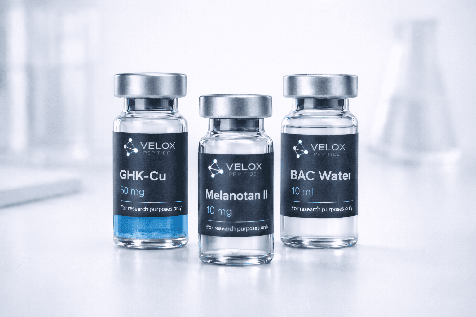
Compuesto de investigación · Solo para uso científico
Aesthetic Skin Stack — Melanotan II + GHK-Cu Research Formulation
Melanotan II (α-MSH analogue) + GHK-Cu (glycyl-L-histidyl-L-lysine copper) · Combined lyophilised · ≥99% HPLC
- ≥99% purity for both peptide components
- HPLC-verified with certificate of analysis
- GHK-Cu: peak activity at ~1 nM concentration
- For in vitro and preclinical research only
The Broader GHK-Cu Research Landscape
The new collagen study sits within a larger research programme that has expanded considerably in the past decade. GHK-Cu was once primarily a dermatological interest — cosmetic industry use in creams and serums, which actually hindered its uptake in serious research (anything that's also in face cream gets taken less seriously by basic researchers). But the underlying biology has gradually attracted more rigorous attention.
Genome-wide studies of GHK-Cu's gene expression effects published by Pickart's group in the 2010s found that it significantly modulates expression of hundreds of genes involved in inflammation, collagen metabolism, DNA repair, and cellular senescence. That breadth of action suggests GHK-Cu is doing something biologically fundamental — possibly related to its role as a copper chaperone and its interaction with copper-dependent enzymes including lysyl oxidase (which crosslinks collagen fibres) and cytochrome c oxidase (the terminal electron acceptor in mitochondrial respiration).
The copper chemistry is genuinely interesting and somewhat underexplored. The Gly-His-Lys tripeptide portion of GHK-Cu has high affinity for copper(II) ions, and copper is a cofactor for several enzymes critical to connective tissue biology. Whether GHK-Cu's biological activity is primarily peptide-mediated, copper delivery-mediated, or a synergistic combination of both is an open mechanistic question.
GHK-Cu Research Applications by System
| Research Area | Key Effect | Evidence Level |
|---|---|---|
| Collagen synthesis (fibroblast) | COL1A1/3A1 upregulation via TGF-β/Smad | Moderate — new study adds to existing data |
| MMP-1 / matrix remodelling | MMP-1 suppression, net collagen protection | Moderate — multiple studies |
| Wound healing (in vitro) | Fibroblast migration, angiogenesis (VEGF) | Moderate — cell culture and rodent |
| Hair follicle biology | VEGF, KGF in dermal papilla cells | Preliminary — cell culture data |
| Anti-inflammation | NF-κB suppression, cytokine reduction | Preliminary — in vitro only |
| Copper enzyme biology | Lysyl oxidase, ceruloplasmin interactions | Theoretical — mechanistic hypothesis |
Limitations of the In Vitro Model
The new fibroblast study is well-designed and adds genuine mechanistic insight. But it's worth being clear about what in vitro fibroblast studies can and can't tell you. Monolayer fibroblast cultures are a simplified system — they lack the three-dimensional extracellular matrix environment of actual skin, the vascular supply that GHK-Cu would encounter in tissue, and the complex cell-cell interactions between fibroblasts, keratinocytes, immune cells, and endothelial cells that characterise the dermis.
Collagen production measured in a monolayer culture doesn't automatically translate to improved collagen organisation in tissue, let alone clinically meaningful skin quality changes. The collagen synthesis finding is mechanistically relevant — it tells you something real about what GHK-Cu does to fibroblast biology. But the gap between "upregulates COL1A1 mRNA 2.3-fold in culture" and "demonstrably improves skin structure in humans" is large and requires its own investigation.
The research roadmap from here would include three-dimensional skin equivalents or organotypic cultures, ex vivo human skin models (which better preserve the full tissue architecture), and eventually properly controlled topical or systemic administration studies in animal models. The in vitro data provides the mechanistic rationale for those more complex studies — which is exactly what it should do.
Research Design Note: 3D Culture Systems
Researchers seeking to advance beyond monolayer fibroblast assays should consider commercially available reconstructed human dermis models (e.g., SkinEthic HDF, LONZA NHDF in 3D) or organotypic skin equivalents combining fibroblasts in collagen gels with keratinocyte overlayers. These systems better capture collagen matrix organisation and allow assessment of whether GHK-Cu's fibroblast-stimulating effects translate into measurable changes in extracellular matrix architecture — a more physiologically relevant endpoint than mRNA upregulation alone.
GHK-Cu in Aesthetic Research Context
For researchers working in aesthetic biology and skin science, GHK-Cu sits at the intersection of several converging research areas: collagen biology, melanocortin signalling (relevant to the companion compound Melanotan II), wound healing, and cellular senescence. The convergence of these areas in dermal research makes the Aesthetic Skin Stack (Melanotan II + GHK-Cu) a useful combined research tool for laboratories studying both the pigmentation and structural aspects of skin biology simultaneously.
Melanotan II (MT-2) is a synthetic α-MSH analogue that activates melanocortin receptors, driving melanogenesis and photoprotective pathways. Combined research with GHK-Cu allows investigators to study collagen synthesis and melanin production in the same cellular system — relevant for understanding the integrated biology of photoageing, where both pathways are affected simultaneously.
Research use only. GHK-Cu and the Aesthetic Skin Stack are not approved for human therapeutic or cosmetic use. VeloxPeptide supplies research-grade peptides for in vitro and preclinical laboratory research with ≥99% HPLC purity certification.